HDR imaging platform
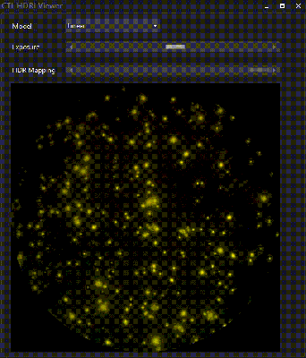
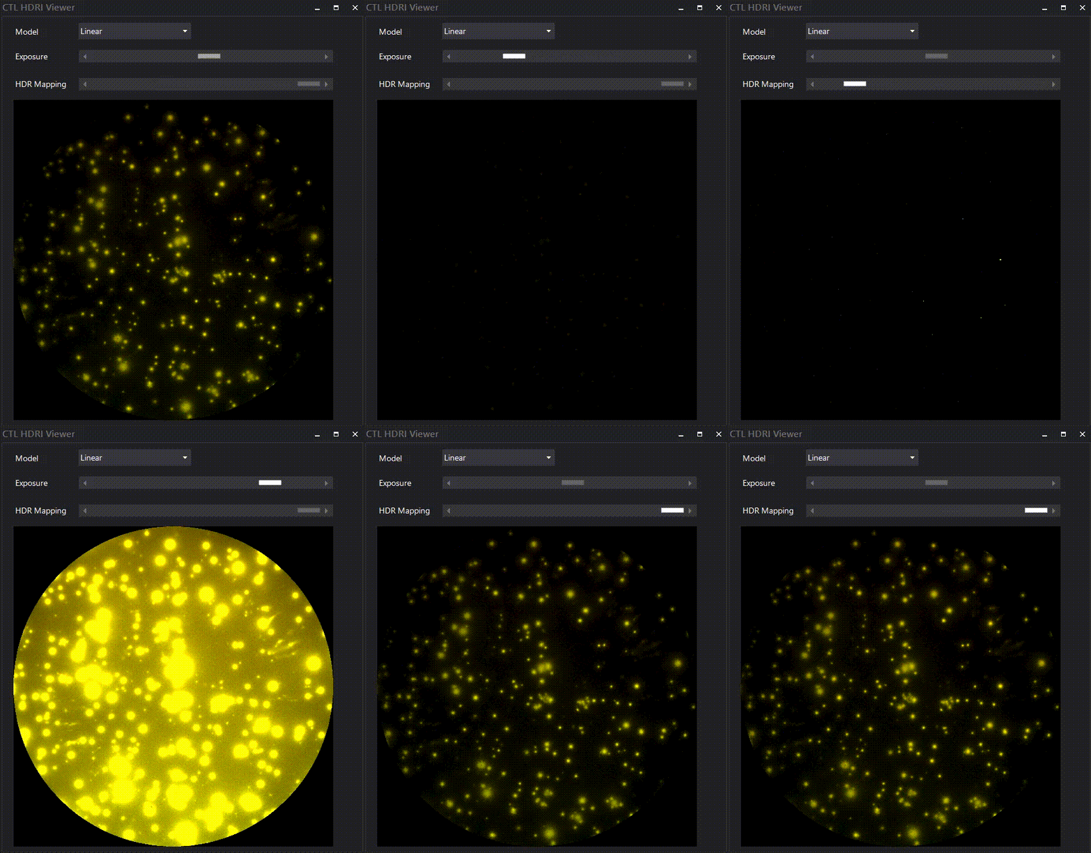
ImmunoSpot® HDR images are generated during the scanning process using multiple different exposures (a process called “bracketing”) for each channel of each well in a FluoroSpot plate. Though these HDR images cannot be displayed in native format on the monitor, the Virtual Exposure Control built into the CTL HDR viewer enables the visualization of strong and weak spots in the same composite image in the counting software – to visually resolve tight spot clusters, and help determine whether cell movement has caused a series of spots, or spot trails.
The additional HDR Image Mapping Control highlights the spot intensity peaks detected at different virtual exposure levels – facilitating visual confirmation and control over software adjustments for accurate multi-color analysis of closely situated spots, in the same channel as well as in different channels for detection of polyfunctional cells secreting multiple cytokines simultaneously.
CTL’s fluorescence-enabled ImmunoSpot® analyzers support multiple high-resolution image formats for optimizing both the speed and accuracy of FluoroSpot counting applications. For high-throughput multi-color counting, where quick turn-around of assays generating large amounts of data are limiting factors, one should consider whether it is truly necessary to scan the images with the highest possible resolution, as this results in larger image file sizes, and takes longer to process each image. A color resolution of 8 bit RGB images (256 levels of intensity per channel) are generally more than adequate for accurate multi-channel FluoroSpot assay analysis. For high-content quantitative analysis of multi-color FluoroSpot data, RAW 16 bit images (65,536 levels of intensity per channel) is an option available in the Fluoro-X image scanning settings.


