FCS output
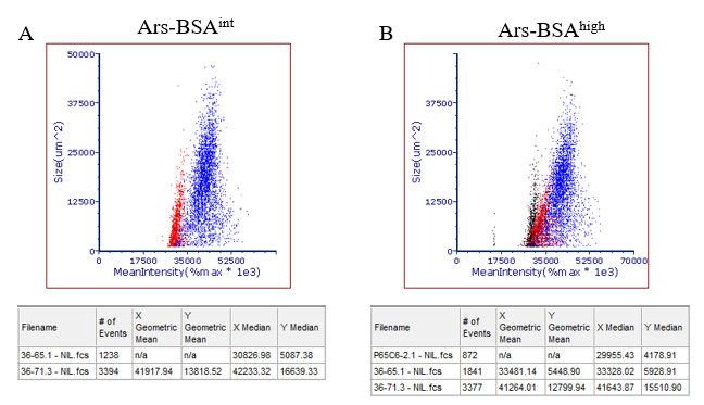
Figure Legend:
Clonal B cell hybridoma lines secreting monoclonal antibody (mAb) with low- (P65C6-2.l, black dots), intermediate- (35-65.1, red dots) or high-affinity (36-71.3, blue dots) for a model antigen (p- azophenylarsonate = Ars) were assessed for FluoroSpot formation. FluoroSpots generated in wells coated with A) Ars-BSAint or B) Ars-BSAhigh were revealed using the Murine IgGl Single-Color FluoroSpot detection system and counted using ImmunoSpot 7 software. Counted FluoroSpots from replicate wells were then merged into a flow cytometry standard (FCS) output. Differential affinity of the anti-Ars mAb was confirmed through plotting the mean intensity and size of individual Fluorospots generated from the respective B cell hybridomas.